CHLAMYDOMONAS GUIDE A model organism for the life sciences
What isChlamydomonas?
Chlamydomonas reinhardtii
A small green alga with a Nobel Prize connection.
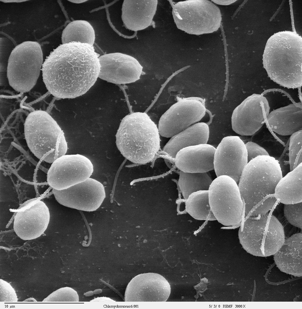
Research into how this alga senses light and swims helped establish optogenetics, a way to study nerve cells using light. From photosynthesis, cilia and reproduction to metabolism and biotechnology, Chlamydomonas lets researchers investigate many aspects of life in a single cell, supporting work across the life sciences.[2][7]
01 — FROM ALGAE TO A NOBEL PRIZE
From a small green alga to a Nobel Prize.
2026 Nobel Prize in Physiology or Medicine
For their discoveries concerning light-gated ion channels and optogenetics, Karl Deisseroth, Peter Hegemann and Georg Nagel were named the recipients of the 2026 Nobel Prize in Physiology or Medicine. One starting point for this work was the study of how Chlamydomonas responds to light.[7]
Discovered in Chlamydomonas, channelrhodopsin is a protein that forms an ion channel that opens in response to light. Introducing this protein into nerve cells to control their activity with light helped establish optogenetics. Basic research into the behavior of a tiny green alga thus led to tools for understanding how neural circuits work.[8][9]
Light sensing is only part of the story. Chlamydomonas also lets scientists study photosynthesis, flagellar movement, reproduction, cell division and metabolism in the same organism. This page introduces that breadth of research.[2][3]
Read the U.S. National Science Foundation explanation02 — BASICS
What kind of organism is it?
Chlamydomonas is a single-celled green alga with a nucleus and a chloroplast. “Single-celled” means that one cell is a complete organism. It uses light to make organic compounds, swims by moving its flagella, and reproduces by cell division.[1]
Size of the cell body
About one hundredth of a millimeter. Size varies with the strain and the state of the cell.
Flagella for swimming
They extend from the front of the cell and move together.
One large chloroplast
A cup-shaped chloroplast occupies a large part of the cell.
The size and structures described here mainly refer to the widely studied species C. reinhardtii.[2]
Where does it live in nature?
Members of the genus Chlamydomonas occur in habitats including fresh water and damp soil. C. reinhardtii includes lineages isolated from soil, and many commonly used laboratory strains descend from soil isolates. Research continues to connect its life in nature with its properties in the laboratory.[1][3]
03 — WHY CHLAMYDOMONAS?
One model organism, many questions about life.
An organism widely used by scientists to investigate how life works is called a model organism. With Chlamydomonas reinhardtii, researchers combine cultivation, genetics, biochemistry and microscopy to study everything from molecular functions to cell behavior.[2][3]
Vary the growth conditions
Researchers can adjust light and nutrients and compare how cells respond. With acetate as a carbon source, cells can grow even in darkness, making it possible to study strains with defects in photosynthesis.[1][2]
Use genetics and crosses
During normal vegetative growth, cells are haploid: they carry one set of chromosomes. Mutants, gene introduction, crosses and meiosis can all be used to connect genes with cell traits.[1][2]
Observe, measure and connect
Scientists can observe living cells and flagella and measure photosynthesis, movement, growth and cellular compounds. Comparing changes in structure with changes in function helps test mechanisms.[2][10]
Shared resources include genome information, mutant strains and plasmids. A major strength is the ability to study mechanisms shared with plant photosynthesis and animal cilia in the same single-celled experimental system.[1][2][10]
Comparisons with other organisms must distinguish shared mechanisms from species-specific traits. For example, relating basic ciliary mechanisms studied in Chlamydomonas to the function of human organs also requires validation in other experimental systems.[10]
04 — FIELDS OF RESEARCH
Diverse research fields, starting with one small cell.
Photosynthesis, swimming, light sensing, mating, growth and metabolic changes provide starting points for research across many fields. Scientists choose genes, observations and measurements to match the questions they want to answer.[2][3] The atlas below uses the same eight categories to filter research groups.
CCM and the pyrenoid
How does the cell use carbon in water?
The CO₂-concentrating mechanism (CCM) takes up inorganic carbon and concentrates CO₂ near Rubisco, the carbon-fixing enzyme. Research examines the formation of the Rubisco-rich pyrenoid and how transport and regulation respond to the environment.[5]
Photosynthesis
How is light energy put to work?
Research covers photosynthetic electron flow, protection against excess light, damage and recovery of the photosynthetic machinery, and gene expression in the chloroplast. Growing and comparing mutants with altered photosynthesis helps reveal how photosynthesis is regulated in response to the environment.[2][3]
Flagella and motility
How do cells swim and generate movement?
Chlamydomonas flagella share their basic structure with animal cilia, including human cilia. Research examines molecular motors, intraflagellar transport (IFT), length control and regeneration. Following swimming behaviors such as phototaxis—moving toward or away from light—helps explain how cells move and the mechanisms involved in ciliary disorders.[10]
Cell cycle and cell structure
When do cells divide, and how do they build structures?
A grown cell can divide successively to produce two, four, eight or more daughter cells, a process called multiple fission. Varying cell size, light and nutrients helps reveal how growth is coordinated with DNA replication and cell division.[11]
Centrioles that organize flagellum formation, and cytoskeletal components such as microtubules and actin, are also studied. Researchers examine how structures that support cell shape and cilium formation are built and maintained.[10]
Metabolism and biomanufacturing
How are carbon and energy used to make useful products?
Studies cover photosynthesis and respiration, acetate use, responses to nutrients such as nitrogen, and the accumulation of starch and lipids. Chlamydomonas provides a model for testing how carbon and energy are allocated to growth or storage.[3][14]
Research includes engineering metabolism to produce compounds such as lipids, making recombinant proteins, and generating hydrogen under oxygen-poor conditions. Productivity must be considered together with growth and cultivation conditions.[12][13][14]
Genetics and genomics
How is genetic information used and passed to the next generation?
Gametes of the + and − mating types recognize one another, adhere and fuse to form a zygote. During germination, the zygote undergoes meiosis and produces haploid offspring. This life cycle and genome information are used to study nuclear inheritance, the uniparental inheritance of chloroplast and mitochondrial DNA, and the regulation of gene expression by small RNAs such as microRNAs.[1][3]
Environmental responses and biological rhythms
How do cells respond to environmental change and anticipate day and night?
Research examines how cells respond to changes in light, gravity, touch, temperature and nutrient availability. Photoreceptors, ion channels, intracellular signals and coordinated flagellar movements can be followed within a single cell to reveal how stimuli change behavior and physiology. Work on channelrhodopsins also grew from studies of light sensing.[3][4][8]
Circadian clocks, which generate rhythms of roughly 24 hours and support adaptation to day and night, are another research focus. Comparisons with other green algae help reveal the functions of clock genes and proteins, as well as the evolution of these clocks.[2]
Evolution and diversity
What do single-celled and multicellular organisms share?
Comparing Chlamydomonas with related volvocine algae and other green algae reveals shared and distinct features of cell shape, movement and life cycles. These comparisons provide experimental systems for investigating the evolution of multicellularity and changes in responses to light that accompany it.[2]
Explore labs in Japan
Open full mapFind research groups and strain resources by location, research field or keyword. The map interface and lab descriptions are in Japanese.
05 — CELL STRUCTURE
What is inside the cell?
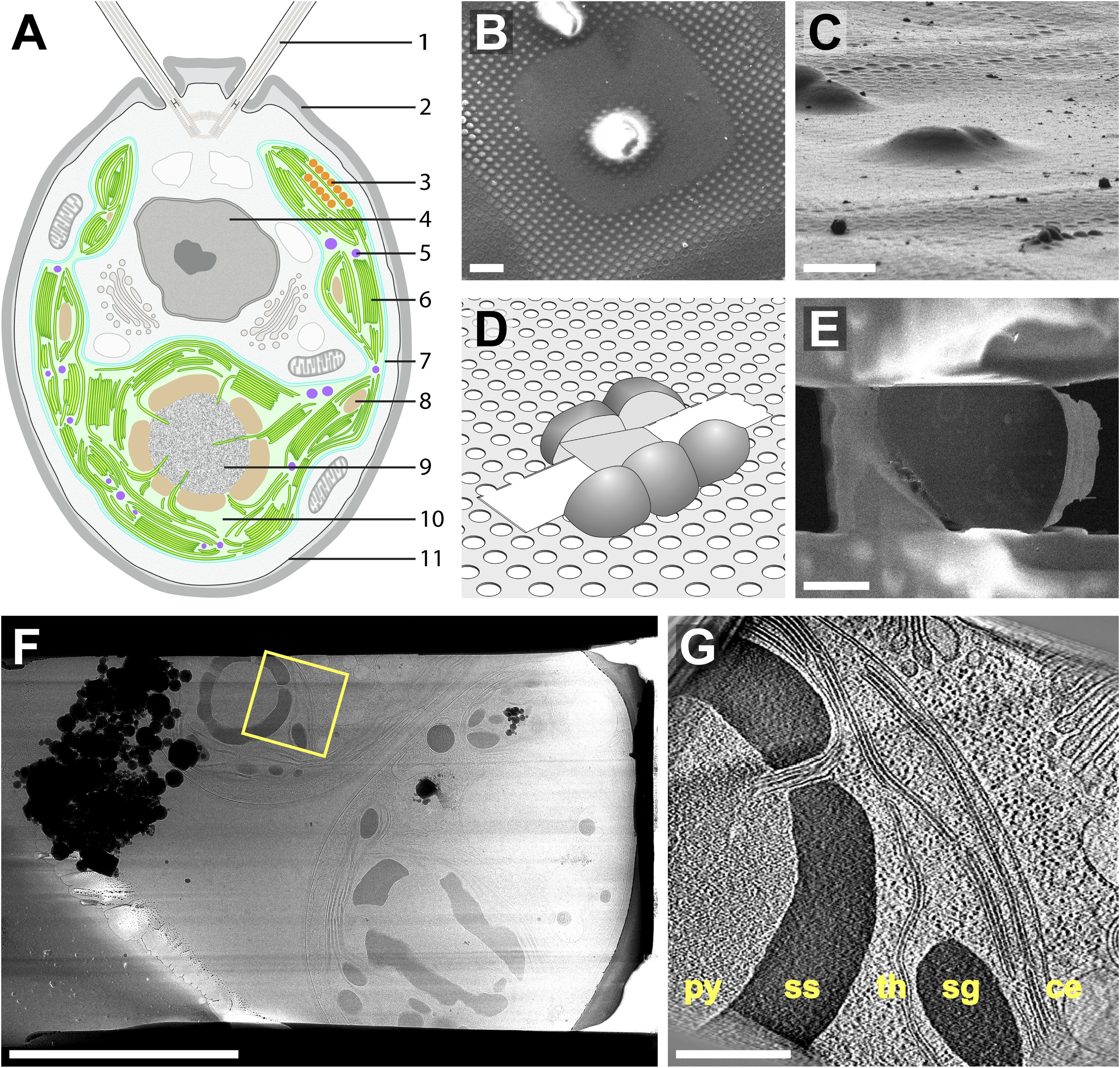
Flagella enable swimming, the chloroplast performs photosynthesis, and the nucleus holds genetic information. These and other structures work together in a single cell. The schematic and tomographic image below come from the cryo-electron tomography study by Engel and colleagues (2015).[6]
- Flagella and cilia
- These structures propel the cell through water. They share their basic structure with motile cilia in animals.
- Chloroplast
- This organelle uses light energy for photosynthesis. It contains the pyrenoid and membranes that carry out photosynthetic reactions.
- Nucleus
- The nucleus contains many genes, holding information needed for the cell’s activities.
- Eyespot
- The eyespot helps the cell detect the direction of light. Pigment layers inside the chloroplast work with photoreceptors in the nearby plasma membrane.
- Mitochondria
- Mitochondria carry out respiration and other metabolic processes, contributing to energy use and the exchange of compounds within the cell.[2][3]
- Pyrenoid
- The pyrenoid is a structure within the chloroplast where Rubisco, the CO₂-fixing enzyme, concentrates. It supports carbon fixation at the center of the CCM.
Take a closer look under the microscope.
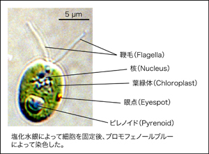
This light micrograph shows a fixed and stained cell. Compare it with the schematic to locate several structures within the small cell. The scale bar is 5 µm.
06 — LIGHT AS ENERGY & INFORMATION
Light provides both energy and information.
Light as energy: photosynthesis
Photosynthesis in the chloroplast uses light energy to incorporate CO₂ into organic compounds. Researchers study proteins that transfer electrons and mechanisms that dissipate excess light energy. Supplying acetate allows strains with photosynthetic defects to grow, helping identify which genes are required for each process.[2][3]
Light as information: phototaxis
At the eyespot, pigment layers inside the chloroplast work together with light-sensitive proteins in the nearby plasma membrane. Signals from light reception alter flagellar movement and change the direction of swimming. Movement in relation to the direction of light is called phototaxis.[3][4]
Depending on the conditions, cells can swim toward or away from light. The eyespot does not form images of the surroundings; it is part of a system that guides movement in response to light direction. Researchers distinguish the use of light as energy from its use as information, and also investigate how the two are connected.[3]
07 — OUR RESEARCH
Research in the Yamano Group
Within this broad field, the Yamano Group studies the pyrenoid and CO₂-concentrating mechanism that support photosynthesis. Using genetics, imaging and physiological analysis, we investigate how molecules assemble into reaction compartments, how their functions are regulated by the environment, and how their structures are inherited through cell division.
08 — QUESTIONS
Common questions
Is it a plant or an animal?
Chlamydomonas is a green alga. It shares photosynthetic mechanisms with land plants, while its flagella share their basic structure with animal cilia. This combination makes it useful across plant science and cell biology.[2][10]
Can it grow in the dark?
C. reinhardtii can grow in darkness when supplied with acetate as a carbon source. Varying light and carbon sources makes it possible to study relationships among photosynthesis, respiration and other metabolic processes.[1][3]
How do cells reproduce?
Cells normally reproduce by cell division. In multiple fission, a grown cell divides successively to produce several daughter cells. They also reproduce sexually: gametes of the + and − mating types fuse, and the resulting zygote undergoes meiosis during germination.[1][11]
Can it help us understand human cilia?
Many components of flagella and cilia, their internal transport systems and their movement mechanisms are shared with humans. In Chlamydomonas, these basic mechanisms can be studied by comparing mutants and observing flagellar regeneration.[10]
How is it used in fuel and protein research?
Scientists study lipid accumulation and hydrogen generation under oxygen-poor conditions, and introduce genes to produce proteins of interest. Work toward applications requires understanding how these products are made and how yield relates to cell growth and cultivation conditions.[12][13][14]
09 — REFERENCES
References and related links
This guide draws on official resources and scientific literature to explain the organism and its research uses. The following sources provide further detail.
- Chlamydomonas Resource Center. About Chlamydomonas. Official guide from the resource center ↗
- Salomé PA & Merchant SS (2019) A Series of Fortunate Events: Introducing Chlamydomonas as a Reference Organism. The Plant Cell 31, 1682–1707. Review of its biology and research history ↗
- Sasso S, Stibor H, Mittag M & Grossman AR (2018) From molecular manipulation of domesticated Chlamydomonas reinhardtii to survival in nature. eLife 7, e39233. Review of its natural history and research uses ↗
- Mittelmeier TM et al. (2013) Independent localization of plasma membrane and chloroplast components during eyespot assembly. Eukaryotic Cell 12, 1258–1270. Research article on eyespot composition and assembly ↗
- Yamano T (2025) Algal Pyrenoids in Global Carbon Cycling: Frontier Research on Structure, Function, and Convergent Evolution. Algal Science and Technology 18, 13–25. Review by our group (PDF, Japanese) ↗
- Engel BD et al. (2015) Native architecture of the Chlamydomonas chloroplast revealed by in situ cryo-electron tomography. eLife 4, e04889. Research article with the cell schematic and cryo-tomograms ↗
- U.S. National Science Foundation (October 14, 2026). NSF BIO congratulates winners of the 2026 Nobel Prize in Physiology or Medicine. The award and its connection to Chlamydomonas ↗
- Nagel G et al. (2003) Channelrhodopsin-2, a directly light-gated cation-selective membrane channel. PNAS 100, 13940–13945. Original research on a light-gated channel ↗
- Boyden ES et al. (2005) Millisecond-timescale, genetically targeted optical control of neural activity. Nature Neuroscience 8, 1263–1268. Original research on optogenetics ↗
- Marshall WF (2024) Chlamydomonas as a model system to study cilia and flagella using genetics, biochemistry, and microscopy. Frontiers in Cell and Developmental Biology 12, 1412641. Review of Chlamydomonas as a cilia research model ↗
- Cross FR & Umen JG (2015) The Chlamydomonas cell cycle. The Plant Journal 82, 370–392. Review of the cell cycle and multiple fission ↗
- Melis A et al. (2000) Sustained photobiological hydrogen gas production upon reversible inactivation of oxygen evolution in the green alga Chlamydomonas reinhardtii. Plant Physiology 122, 127–136. Research article on hydrogen production under defined conditions ↗
- Rasala BA et al. (2010) Production of therapeutic proteins in algae, analysis of expression of seven human proteins in the chloroplast of Chlamydomonas reinhardtii. Plant Biotechnology Journal 8, 719–733. Research article on recombinant protein production ↗
- Boyle NR et al. (2012) Three acyltransferases and nitrogen-responsive regulator are implicated in nitrogen starvation-induced triacylglycerol accumulation in Chlamydomonas. Journal of Biological Chemistry 287, 15811–15825. Research article on nitrogen deprivation and lipid accumulation ↗