Our Research Philosophy
How does life form organized reaction compartments on its own, maintain them, and pass them on to the next generation? This is the question that guides our research. A "reaction compartment" is a space where the required molecules gather and specific chemical reactions take place. At the heart of our work is the link between how molecules come together and how these assemblies keep working to support life.
Using the pyrenoid in microalgae as a model, we study "phase separation," a process in which molecules gather into liquid-like droplets. Like the separation of oil and water, this process creates compartments without membranes, called "membrane-free organelles." Unlike equilibrium systems that move toward a stable state, living reaction compartments work in a nonequilibrium state, with raw materials entering, products leaving, and energy being used continuously.
These compartments replace their molecules and change their shape while maintaining their function and passing it on through cell division. We study formation, maintenance, and inheritance as one continuous process to understand how flexibility and stability work together. We also ask which environments favor reaction compartments and when they are lost, extending our view from the life of a cell to the history of evolution. By linking molecules, functions, and environments, we seek common principles of how life creates order and adapts to change.

Pyrenoid Dynamics -Learning from a Changing Reaction Compartment-
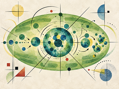
We explore this question using Chlamydomonas, a single-celled green alga. Its chloroplast contains the pyrenoid, a reaction compartment in which Rubisco, the enzyme responsible for photosynthetic carbon fixation, gathers at high density. Its core forms through phase separation and works with the surrounding starch and the membranes that pass through it. This model lets us connect changes in structure and movement to a measurable function: how well the cell uses carbon [1].
We showed that LCIB, a protein needed for carbon use, moves to the region around the pyrenoid in response to CO2 levels. We also found that the surrounding starch structure supports both LCIB positioning and the CO2-concentrating mechanism. The pyrenoid is not a static mass of enzymes. It is a reaction compartment that rearranges its components in response to the environment [2,3,4].
We now combine live imaging, which follows living cells over time, with machine learning to study cells before, during, and after division. In a submitted study, we detected easily overlooked shape abnormalities and linked them to defects in rebuilding the pyrenoid during cell division and to CO2-dependent growth. By connecting shape, movement, and function, we aim to understand how reaction compartments pass their function on to the next generation [5].
[1] Yamano T (2023) Plant Morphology 35, 3-8.
[2] Yamano T et al. (2010) Plant Cell Physiol 51, 1453-1468.
[3] Yamano T et al. (2022) Plant Physiol 188, 1081-1094.
[4] Toyokawa C et al. (2020) Plant Physiol 182, 1883-1893.
[5] Matsuo K and Yamano T. bioRxiv
How the CCM Works -Bringing Carbon from Water to the Reaction Compartment-
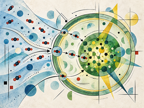
Photosynthesis requires not only enzymes but also a supply of their raw material, CO2. In water, limited CO2 supply can restrict photosynthesis. Many algae therefore take up carbon from their surroundings and concentrate CO2 around Rubisco using the CO2-concentrating mechanism (CCM). The pyrenoid carries out carbon fixation at the center of this system.
We have studied how carbon in the form of bicarbonate ions is transported from outside the cell into the chloroplast. We showed that HLA3, a transporter in the plasma membrane, and LCIA, a transporter in the chloroplast envelope, work together to support carbon uptake. Transport across these two boundaries forms a single coordinated pathway [6].
We also showed that CAH1, an extracellular enzyme that speeds up the conversion between CO2 and bicarbonate, helps cells use carbon efficiently, especially when CO2 is scarce relative to bicarbonate. Carbon uptake requires not only transport across membranes, but also conversion into a chemical form that cells can readily take up [7].
[6] Yamano T et al. (2015) PNAS 112, 7315-7320.
[7] Shimamura D et al. (2024) Plant Physiol 196, 2395-2404.
CCM Regulation -Turning It On When Needed and Off When Not-
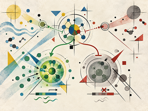
The CCM is a powerful carbon-collecting system, but it requires energy. Cells use their limited energy efficiently by increasing CCM activity when CO2 is scarce and reducing it when it is abundant. We study this switching process through gene expression, which determines when and how much of each protein is made.
We showed that signaling through the chloroplast protein CAS is needed for the expression of nuclear genes encoding carbon transporters. Losing SAGA1, a protein involved in pyrenoid structure, disrupts CAS positioning and the gene expression it controls. Based on these findings, we propose that pyrenoid structure is linked to the regulation of carbon uptake across the cell [8, 9].
We discovered CBP1, a nuclear protein that represses the CCM. When CO2 is abundant, CBP1 reduces the expression of CCM-related genes. We also identified the cytosolic factor HCR1 as a second repressor that helps shut down the CCM. These repressors, which act in the nucleus and the cytosol, are helping us understand how cells control CCM activation and shutdown [10, 11, 12].
[8] Wang L et al. (2016) PNAS 113, 12586-12591.
[9] Shimamura D et al. (2023) Photosynth Res 156, 181-192.
[10] Shimamura D et al. (2026) PNAS 123, e2518136123.
[11] Shimamura D and Yamano T (2026) Front Plant Sci 17:1882169.
[12] Miichi S et al. bioRxiv
Technology Development -Turning Overlooked Cell Differences into New Discoveries-
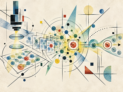
Small differences in cell shape may hold clues to how reaction compartments work. Finding rare abnormalities among many cells, isolating those cells, and studying them in detail requires tools for observation, analysis, and handling to work together. We develop these tools to answer biological questions.
Working with researchers in engineering, information science, and other fields, we helped develop AI-based image-activated cell sorting. This technology analyzes microscope images in real time and selects living cells with the features of interest [13, 14].
We also developed a method for introducing genes into Chlamydomonas without removing the cell wall, providing a basis for studying gene function [15]. By combining these methods with live-cell observation and machine-learning analysis of cell shape, we will test which genes and molecular functions explain the differences we find.
We use AI to find differences, microscopy to follow changes, and genetic tools to test mechanisms. We keep technology development closely tied to our biological questions, building better ways to understand how reaction compartments form, work, are regulated, and are passed on.
[13] Nitta N et al. (2018) Cell 175, 266-276.
[14] Nitta N et al. (2020) Nat Commun 11, 3452.
[15] Yamano T et al. (2013) J Biosci Bioeng 115, 691-694.